
Technology refined by dedicated surgeons.
In collaboration with radiologists and surgeons, Moventis Imaging has built a CT-compatible setup with paired image acquisition and structured reporting, giving teams an additional mechanical view in painful or unclear knee replacement follow-up.
 01
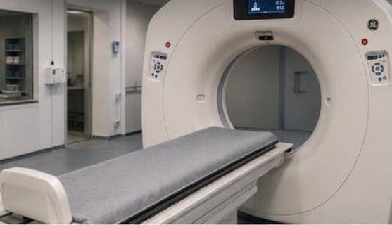
01Existing CT scanner
No additional scanner needed. Moventis works with standard clinical CT equipment already in the department.
 02
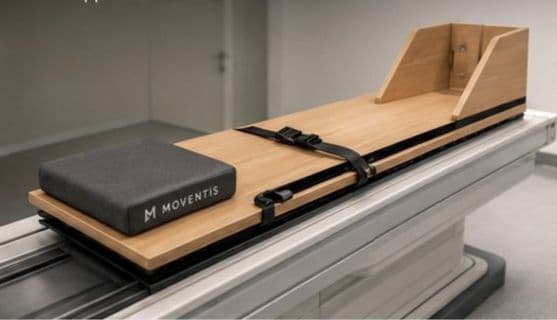
02Moventis device (illustrative)
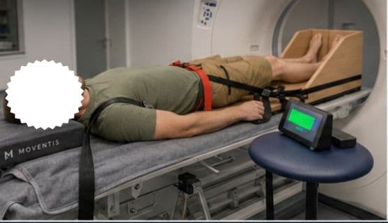
A loading board that reproduces knee loading conditions under body weight, fully compatible with existing CT scanners, without requiring the patient to stand.
 03
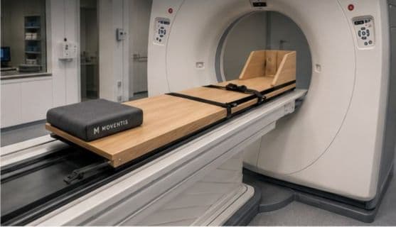
03Device placed on CT table
Quick setup and alignment. The device positions directly on the standard CT table with no modification to the scanner.
 04
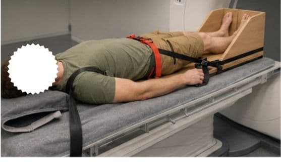
04Patient positioned
The knee is aligned near the gantry isocenter. An unloaded scan is taken first.
 05
05Controlled load and acquisition
A second scan is taken in the same position, while the patient engages the device enabling acontrolled axial load.
 06
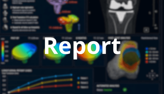
06From images to structured output
Automated analysis, visualisation, and reporting. The two scans are compared to generate structured visual and numerical outputs for review and discussion.
Mimicking weight-bearing, without asking the patient to stand
Standard CT is acquired at rest. But the knee spends most of its time under load, and some mechanical changes only become apparent when compressive forces are present. The Moventis setup applies a controlled axial load while the patient lies supine inside the scanner, reproducing the compressive conditions of weight-bearing without requiring the patient to stand.
This makes it possible to compare implant behaviour between an unloaded and a loaded condition in a single session, with no repositioning between scans. Better acquisition consistency means a more interpretable comparison.
Protocol considerations include consistent positioning, paired scan comparability, metal-aware reconstruction, and dose-conscious imaging.
An additional mechanical view, not a standalone verdict
The output is intended to support interpretation and case discussion alongside standard care, not to determine the cause of pain on its own. Measured movement between unloaded and loaded conditions does not automatically indicate loosening or explain symptoms.
Interested in the approach?
We are open to conversations with orthopaedic and radiology teams, as well as implant manufacturers interested in implant behaviour and evidence generation.